Orthopaedic Management & Surgery
What is osteochondritis dissecans?
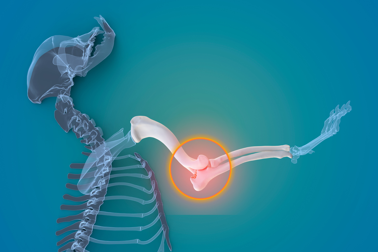
The term osteochondrosis refers to an abnormal development of the cartilage on the end of a bone in the joint. Osteochondritis dissecans (OCD or OD) is an inflammatory condition that occurs when the diseased cartilage separates from the underlying bone. It most commonly affects the shoulder joint but the elbow, hip, or knee (stifle) may also be involved.
Which breeds of dogs are likely to be affected by this condition?
This is a developmental disease that occurs in rapidly growing large breed dogs typically between 6 and 9 months of age and tends to occur more often in male dogs. The cause of OCD is unknown.
However, this disease is more common in dogs receiving too much energy and calcium in the diet. Other factors may also include genetics, rapid growth, trauma, lack of blood flow, and hormonal factors.
What are the signs of OCD?
Dogs that are affected with OCD typically limp or are lame in the affected leg or legs. During an orthopedic examination, when pressure is applied to the affected joint or when the joint is manipulated, the dog will often cry out in pain. The affected joint may be swollen and warm to the touch. In some cases, the lameness may be mild and intermittent while in other cases, the dog may be in constant pain and avoids bearing weight on the affected leg.How is OCD diagnosed?
The results of a lameness examination may be suggestive of this condition, especially if the shoulder is the affected joint. If one of the other joints, such as the stifle (knee), hip, or elbow, other bone conditions must also be considered, including hip dysplasia, patellar luxation, and elbow dysplasia.
Because of the possibility of permanent lameness, your veterinarian will recommend diagnostic testing if the lameness persists for more than 2 weeks. Radiographs (X-rays) are usually performed to investigate lameness. Several radiographs of each affected leg are necessary in order to get an accurate assessment of various bones and joints. In many cases, this will require a short-acting anesthetic or sedative in order to achieve the optimal positioning for diagnostic purposes. In dogs under 6-7 months of age, X-rays can be challenging to interpret due to the presence of growth physes (growth plates). To reach the diagnosis, it may be necessary to have the X-rays examined by a veterinary radiologist.
In some cases, an arthroscopic examination may be required to reach or confirm the diagnosis.
How is OCD treated?
The OCD lesion can vary in severity, ranging from a crack in the cartilage, to a cartilage flap, to a completely detached fragment of cartilage that is floating around in the joint (called a joint mouse).
If the defect is a crack or a very small flap of cartilage, it may heal if the patient has strict rest and activity restrictions for several weeks. In these cases, the dog will be restricted to short leash walks only, and cage rest will be strongly recommended. Medications to relieve inflammation and supplements to promote joint health will usually be prescribed. Often, you will be taught how to perform passive range of motion (PROM) exercises, in which you will move the joint through its normal range of motion while your dog is lying on his side. This is done to maintain the joint's flexibility and mobility.
If the lameness does not improve following this conservative approach, if the cartilage flap becomes folded in the joint, if the cartilage defect is large, or if a piece of cartilage breaks free, surgery will be required to remove the defective flap or the floating piece of cartilage. This may be done by surgically opening the joint or by using an arthroscope. Whatever the surgical technique, the remainder of the cartilage surface will be inspected and any other areas of defective cartilage will be debrided or removed.
What sort of aftercare will my dog require following surgery?
Surgical removal of the diseased cartilage will relieve the inflammation and pain, allow the joint surface to remodel, and minimize the development of degenerative joint disease.
For the first 2-3 weeks postoperatively, your dog will be restricted to short leash walks only, and you will be instructed on how to perform PROM exercises. Anti-inflammatory medications and joint supplements will be prescribed. After 3 weeks, the amount of controlled exercise will be increased and specific rehabilitation exercises such as swimming may be prescribed. After 6 weeks, your dog may progress to other controlled activities such as light jogging.
What is the prognosis following surgery?
The prognosis varies depending on the joint that is affected. If the shoulder joint is affected, the prognosis is good; if the elbow joint is affected the prognosis is guarded. In all cases, the prognosis improves if surgery is performed early in the course of the disease, before secondary degenerative joint disease occurs.
Weight control is important to avoid unnecessary stress on the joint.


