Conjunctival Graft for Rupture Globe
What is globe rupture?
A ruptured globe is an eyeball with full-thickness defect in part of the eye’s wall. It is a serious injury. The eye is blind, very painful, and difficult to be repaired.
The most common causes for globe rupture include neoplasia (cancer around or inside the eye), trauma (such as a severe scratch or puncture to the eye, hitting the eye on something sharp), deep corneal ulceration, and glaucoma (high pressure inside the eye).
Having a ruptured globe will be extremely painful and it will distress your pet. It is an emergency and it should be addressed through surgery as soon as possible to prevent serious complications.

What is a conjunctival graft?
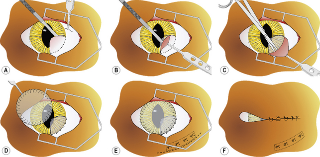
Placing a conjunctival graft is commonly advised for ruptured globe associated with deep corneal ulceration. The conjunctiva is a thin, elastic tissue with great blood supply. When a conjunctival graft is placed, conjunctival tissues are harvested and placed over the defect.
The two main types of conjunctival graft are:
- The conjunctival pedicle or rotational graft: It is commonly used when there is a localised deficit in the cornea. The graft will deliver nutrients and provide structural support. The graft can be trimmed later to reduce the impact on vision.
- The 360-degree conjunctival graft: It is commonly used when the defect is a large area or a central lesion. Significant scar tissues after the operation are expected due to the large area of defects.
What is expected from a conjunctival graft?
After a conjunctival graft is placed into the defect, the conjunctival tissue can provide blood supplies, nutrients and structural support to the ulcer bed and replace the tissue lost to corneal infection when it integrates into the corneal tissue.
The graft may block the vision of the eye and leave a significant amount of scar tissues. If the ulcer is very deep or infected, the prognosis is guarded. Nonetheless, it is more likely than medical treatment to save a ruptured globe.
After the eye is healed, the conjunctival tissue should not be removed and it should be left integrated into the cornea. The scarring process may last for several months. The result can range from a small opacity to a dense white corneal scar.
What is the aftercare?
A ruptured globe is very fragile and susceptible to injury. Wearing a rigid E-collar is strongly advised for your pet to prevent head thrashing or self-trauma.
Topical antibiotics will be administered until the graft has integrated into the ulcer and the eye is healed. The site of the ulcer should be sampled for culture and sensitivity, guiding the choice of the topical antibiotics.
Analgesics are also essential to prevent pain. Since the cornea has a dense network of nerve receptors, analgesics like opioids will be prescribed for controlling corneal pain.
An eye exam will be required in a few days post-surgery and then every 1-2 weeks to assess the healing of the eye.
After there any complications during or after the procedure?
Conjunctival grafts may fail if the original corneal tissue cells were not adequately debrided (cleaned) from the ulcer site if local infection develops or if surgical technique was poor.
A graft may block the vision of the healing eye. It also leaves more scarring than medical therapy. Although the graft may be able to be trimmed under general anaesthesia after recovery, vision may not return due to the significant scarring.
Too much tension on the graft can prematurely detach the graft from the ulcer site. If the conjunctival graft detaches from the cornea and the ulcer ruptures, enucleation will be needed. Extensive leaking from the graft can also occur.
During the healing process, scar tissues will be formed and they may lead to secondary glaucoma.


