Elbow Dysplasia
What is Elbow dysplasia?
Elbow dysplasia refers to abnormal growth or development of the elbow joint. The elbow joint is made up of 3 bones, the radius, the ulna, and the humerus.
Three different problems can cause elbow dysplasia:

1. A ununited anconeal process (UAP)
is a developmental defect of the anconeal process. The anconeal process is a small piece of bone that is found on the back of the ulna (the longer of the two bones of the forearm) at the rear point of the elbow. Normally, as the dog reaches puberty, the growth plate that is found between the anconeal process and the rest of the ulna closes, fusing the parts of the bone together. When this part of the ulna does not fuse, the elbow joint becomes unstable, causing lameness and pain, and quickly develops degenerative joint disease or arthritis. In some cases, the bone fragment floats freely in the joint, causing further discomfort. Dogs with this disease are lame on the affected leg or legs and they may cry when the elbow is extended. Treatment requires surgery. The results are much better if surgery is done before secondary arthritis affects the joint.2. A fragmented coronoid process (FCP)
is a developmental defect of one of the coronoid processes, two small bony protrusions on the end of the ulna, within the elbow joint. In this condition, the medial or lateral coronoid process develops a fissure or crack and separates from the rest of the bone. This causes pain and joint instability. Unless surgery is done immediately, arthritis will develop in the joint. This condition is likely hereditary and occurs more frequently in large breed dogs, especially the retriever, Rottweiler, and German Shepherd.3. Osteochondritis dissecans (OD or OCD) or osteochondrosis (OC)
is a defect on the smooth cartilage surface within one or more joints. The term osteochondrosis refers to an abnormal development of the cartilage on the end of a bone in the joint, while osteochondritis dissecans refers to a separation of the diseased cartilage from the underlying bone. It most commonly affects the shoulder joint but the elbow, hip, or knee (stifle) may also be involved in young, rapidly growing large-breed dogs. In some cases, the defect is a flap of cartilage or a crack in the cartilage on the end of the bone; if this occurs, the defect may heal with strict rest and restriction of activity for several weeks. In these cases, pain medication, cartilage-protecting drugs and nutraceuticals may also be prescribed. In other cases, a piece of cartilage breaks off and floats freely in the joint (sometimes called a "joint mouse"). This causes pain, which varies from mild, intermittent limping to intense, constant pain. Surgery to remove the defective flap or "floating" piece of cartilage is the recommended treatment in these cases. For further information on this condition, see our handout "Osteochondritis Dissecans".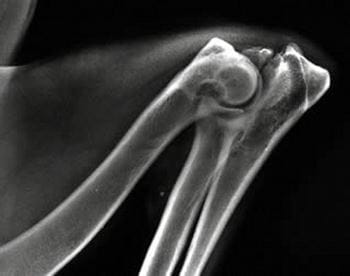
Elbow Dysplasia
For further information on each of the types of elbow dysplasia, contact us for more detail.


